
Forme fruste keratoconus: the stage almost nobody catches in time
Forme fruste keratoconus is the early stage where one procedure can stop the disease. The problem: it rarely gets caught before it is too late.
In short: Forme fruste keratoconus is the early stage where a single procedure can stop the disease. The problem: it does not show up in a normal eye exam. Only one specific study finds it, and many offices do not have it.
There is a version of keratoconus that almost no ophthalmologist catches in a routine visit. It is called forme fruste keratoconus, the stage where the cornea is already changing but has not yet produced clear symptoms. Catching it in time changes the entire course of the disease. It moves from “you can no longer tolerate contact lenses and need rings or a transplant” to “we stabilize the cornea with one procedure and it is done.” The problem is that it only shows up if someone looks for it.
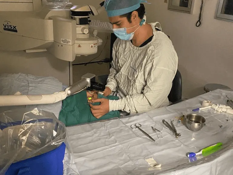
Dr. Emmanuel Bolaños Betancourt: ophthalmic surgeon with subspecialty training in Cornea, Refractive Surgery, and Cataract Microsurgery. I practice at Hospital Aranda de la Parra Puerto Interior (20 minutes from León) and at Oftalmo-Visión Clínica Oftalmológica in downtown Silao. Board certified by the Consejo Mexicano de Oftalmología since 2013. More than 15 years in clinical practice, 292 combined Google reviews with a 4.9 average.
What changes depending on the stage it is caught at
Keratoconus is a disease where the cornea, the clear surface covering the front of the eye, weakens and deforms. It loses its normal round shape and becomes cone-shaped, which distorts vision. Left untreated, the disease progresses: the cornea keeps thinning, keeps deforming, and vision keeps getting worse even if you change your glasses every few months.
Here is what matters most: keratoconus is classified by grade, and treatment changes drastically depending on the grade.
- Forme fruste keratoconus: early topographic changes, patient has no symptoms. Treatable with cross-linking. One procedure stops the disease.
- Grade 1-2: mild symptoms, contact lenses still tolerable. Cross-linking remains the main treatment.
- Grade 3: cornea already significantly deformed. Intracorneal rings in many cases.
- Grade 4: cornea very thin and deformed, visual quality badly compromised. Corneal transplant.
The difference between catching keratoconus at the forme fruste stage and catching it at grade 3 to 4 is the difference between a simple outpatient procedure and major surgery with donor tissue. It is the same disease, at different moments.
That difference between stages is what matters most. And the stage where the most can be done is exactly the one that is hardest to catch.
Why the most treatable stage is the least visible
In a standard eye exam, someone measures your visual acuity, checks your prescription, looks at your eyes with a slit lamp, and examines the back of your eye. None of that catches forme fruste keratoconus. The only way to see it is with two specific studies:
- Corneal topography: a map of the corneal surface. Detects irregularities in curvature.
- Corneal tomography: evaluates internal structure, posterior elevation, and thickness across zones. Detects thinning and bulging that topography alone does not show.
If you have never had these two studies, you cannot assume your cornea is fine, especially if you have any of the warning signs listed below.
If the early stage is invisible on a normal exam, the question is when to actually look for it. There are signs that justify asking for the specific studies.
The signs that justify asking for the study
None of these is conclusive on its own, but together they justify investigating:
- Your prescription changes fast, your glasses get updated every 6 to 12 months, and the new lenses do not fully clear up the blur
- High astigmatism that keeps climbing at each checkup
- Irregular astigmatism, or an axis that shifts between exams
- You rub your eyes a lot, whether from allergies, chronic itching, or dry eye
- A family history of keratoconus
- You were told you are not a LASIK candidate without a clear explanation
- Night vision with halos or glare with no clear cause
- Age between 15 and 30, the highest-risk window for onset
If you have two or more of these, it is worth coming in for an evaluation with corneal topography and tomography. The study takes 15 minutes and gives you a definite answer.
If the studies confirm early keratoconus, there is one procedure that stops it from advancing. And it changed the whole picture since 2003.
Corneal Cross-Linking: what changed in management
Corneal cross-linking (CXL) is an outpatient procedure that strengthens the collagen structure of the cornea. The technique: riboflavin (vitamin B2) is applied to the cornea and activated with ultraviolet light at a specific wavelength. The activated riboflavin creates additional chemical bonds between the collagen fibers, which increases the rigidity of the cornea.
Result: keratoconus progression stops in roughly 95% of cases when treated in time. It does not reverse the deformity that already exists, but it keeps it from advancing.
I use AVEDRO equipment, the international reference for accelerated cross-linking. The accelerated technique cuts the procedure time while keeping efficacy comparable to the original technique.
- Outpatient procedure, no hospital stay
- Numbing drops for anesthesia
- Total duration: 30 to 60 minutes
- A scratchy discomfort for the first 3 to 5 days, controlled with drops
- Functional vision returns in 1 to 2 weeks
- Periodic follow-up with control topographies
When cross-linking is not enough on its own
Cross-linking stops progression, but it does not correct the deformity that already exists. In more advanced keratoconus, there are other complementary or alternative options:
- Specialty contact lenses: rigid gas permeable (RGP), scleral, or hybrid. They improve vision without surgery, but they do not stop progression on their own.
- Intrastromal rings: acrylic segments placed within the thickness of the cornea to regularize its shape. Indicated for grade 2 to 3 with contact lens intolerance.
- Corneal transplant: reserved for grade 4 or cases where the previous options have been exhausted. I perform it with penetrating or lamellar technique depending on the case.
I ran the Corneal Transplant Program at Hospital Regional de Alta Especialidad del Bajío from 2014 to 2017. That transplant experience shapes how I decide when to intervene and with which technique, and just as important, when not to intervene yet.
You now know what it is, why it goes unnoticed, what signs suggest it, and what can be done in the operating room. What is left is the one habit that changes the outlook the most, and almost nobody teaches it. It costs nothing, it happens every day, and almost nobody pays attention to it until it is too late.
The instruction that matters most, and almost nobody follows
Repeated microtrauma from eye rubbing is one of the main causes of keratoconus progression. It is basic physics: every time you rub your eyes, you put pressure on a cornea that is already weakened, and you speed up the deformity.
That is why full keratoconus management includes treating the cause of the itching: allergies, dry eye, chronic conjunctivitis. Stopping the rubbing can matter as much as any corneal procedure.
When does keratoconus stop progressing?
In most cases, keratoconus progresses until age 30 to 35 and then stabilizes on its own. But that is a pattern, not a guarantee. Some patients keep progressing past 35, especially with:
- A strong family history
- Uncontrolled severe allergies
- Chronic eye rubbing
- Hypothyroidism, atopy, or other related systemic conditions
The only way to confirm stability is to compare serial corneal topographies over time. If you have been diagnosed with keratoconus, checkups every 6 to 12 months are not optional.
If you are coming from León or the Bajío region: how the trip works
My surgical office, with the full corneal topography and tomography equipment, is at Hospital Aranda de la Parra Puerto Interior, 20 minutes from León. A complete keratoconus evaluation takes a single visit. If you need cross-linking, we usually schedule it for a second visit. If you need rings or a transplant, we coordinate the dates to minimize your trips.
Last updated: · Reviewed by Dr. Emmanuel Bolaños, ophthalmic surgeon (Mexican Board of Ophthalmology, member 3045).
Want me to look at your case?
Send me a WhatsApp with the reason for your visit. I will tell you whether an evaluation makes sense and which of my two offices to come to, before you make the trip. The whole visit can be in English.